A scanning tunneling microscope (STM) is a type of electron microscope. It is much more powerful than an ordinary microscope of the sort we would use
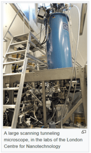
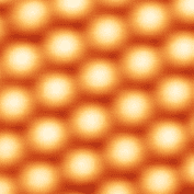
[Image source: O. Usher (UCL MAPS) – http://www.ucl.ac.uk/maps-faculty/potw/potw/potw1401, CC BY 3.0, https://commons.wikimedia.org/w/index.php?curid=30504029 Retrieved from https://en.wikipedia.org/wiki/Scanning_tunneling_microscope July 5, 2017]Silicon atoms on the surface of a crystal of silicon carbide. [Image Source: Guillaume Baffou; GFDL (http://www.gnu.org/copyleft/fdl.html) or CC BY-SA 3.0 (http://creativecommons.org/licenses/by-sa/3.0), via Wikimedia Commons https://en.wikipedia.org/wiki/Scanning_tunneling_microscope Retrieved June 17, 2017]when looking at blood cells or amoebas. An ordinary microscope works on the principle of light, which travels from the cells or amoebas and into our eyes.
Scanning tunneling microscopes allow us to form images of objects as small as atoms, a task not possible with microscopes that rely on light. The STM has a stylus with a tip made of a single atom to probe or “feel” materials atom by atom. The microscope creates an image based on what the probe has felt.
Scanning Tunneling Microscope and Quantum Tunneling
An STM depends on an effect called “quantum tunneling.” Quantum tunneling is the ability of a quantum particle to breach a barrier, even one which, according to the laws of classical physics, it should have insufficient energy to breach. In an STM, electrons tunnel across a gap (the barrier) between the tip of the probe and the surface being examined. Depending on the type of STM, the electrons might be generated by either the tip or the surface being examined. As they tunnel, the electrons generate an electrical signal that is used to form an image of the surface being examined. The animated video to the left shows the quantum tunneling effect and then, how tunneling is put to use. Click image for video.
How the Scanning Tunneling Microscope Works
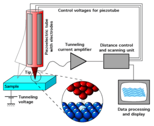
How the scanning tunneling microscope works. [Image source: Michael Schmid – Michael Schmid, TU Wien; adapted from the IAP/TU Wien STM Gallery, CC BY-SA 2.0 at, https://commons.wikimedia.org/w/index.php?curid=180388. Retrieved from https://en.wikipedia.org/wiki/Scanning_tunneling_microscope on July 2, 2017.]The sample being examined is shown as a blue rectangular box. The probe is shown in pink and grey. Its tip is in red. The circular inset shows the red spherical atoms of the tip and blue atoms of the sample. Red dotlike electrons tunnel between the tip and the sample, focusing on a single pale blue atom of the sample. The tunneling electrons create an electric current, which runs through an amplifier. Once amplified, the current feeds a data processing display, which creates an image.
*For simplicity, these have been called “silicon atoms.” However, more accurately, they are ions in a sea of electrons, forming a crystal. Whereas the ions are visible, the electrons are too tiny to show up in the image.

 he animated video to the left shows the quantum tunneling effect and then, how tunneling is put to use. Click image for video.